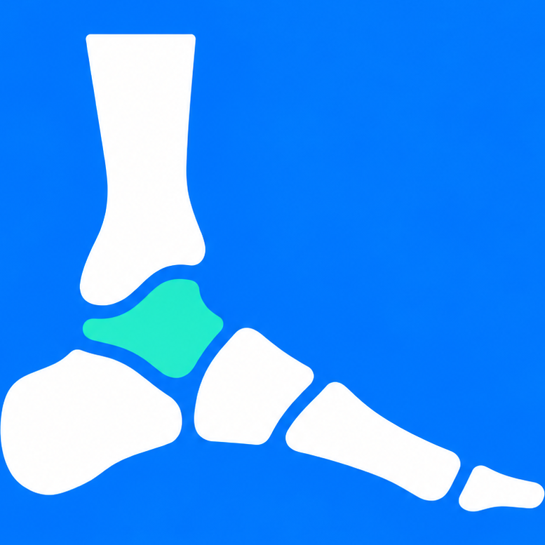
Weight-Bearing CT Is Changing How We Measure the Foot
Three-dimensional imaging under load reveals alignment that plain films and supine CT can miss.
Podiatrify Clinical Editorial Team, Editorial Staff
Podiatric Medicine & Research
- Published
- Published Aug 24, 2026
- Last reviewed
- Last reviewed Aug 24, 2026
- Reading time
- 3 min read
- Audience
- Clinicians
- Evidence level
- Emerging Evidence
Clinical summary
Cone-beam weight-bearing CT gives 3D images of the foot while standing. It is increasingly used in hallux valgus, flatfoot, and hindfoot alignment assessment. Measurement standards are still being built.
Patient view: plain-language reading.
What it adds
Standing 3D imaging shows metatarsal rotation, subtalar alignment, and peritalar subluxation under physiologic load, which supine CT cannot capture.
Limits
Access, cost, and a lack of universally agreed measurement protocols remain barriers. Radiographs are still the first-line study for most foot and ankle complaints.
References
- 1.Weight Bearing CT International Study Group publications. Verify specific papers before citing.
Summaries are editorial. Verify details against the original publications.
Industry Update. Technology and business news. Editorial standards
Podiatrify provides educational information and risk guidance and does not provide a medical diagnosis. Symptoms involving severe pain, rapidly spreading infection, black or blue tissue, major trauma, loss of circulation, or systemic illness may require urgent medical evaluation.